Microbiome Gallery
Explore the visual gallery of microbiome research, from gut bacteria and antibiotics to cellular imaging and gene discovery

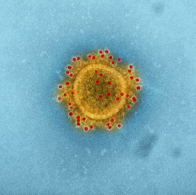
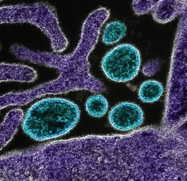
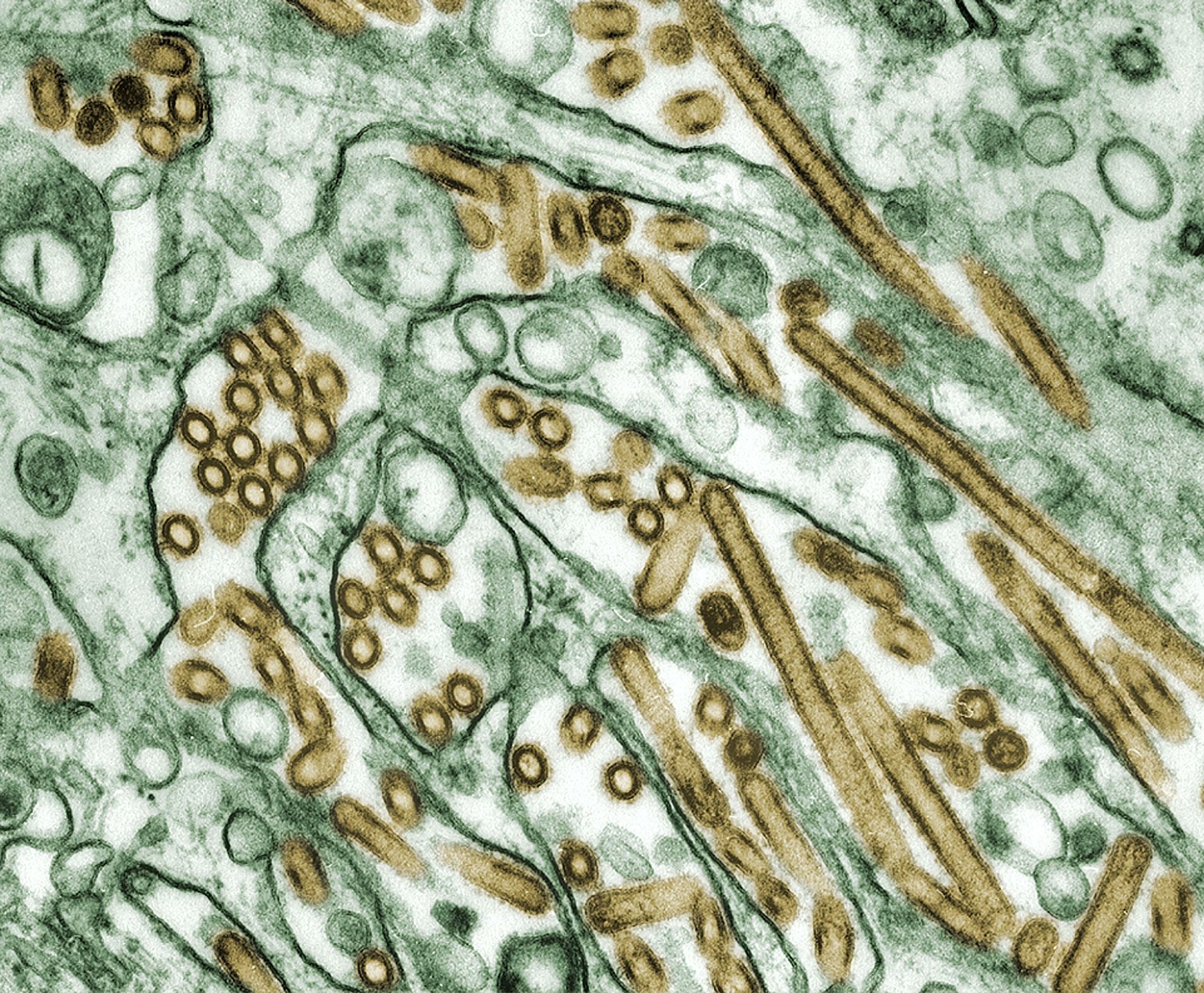
mers-cov
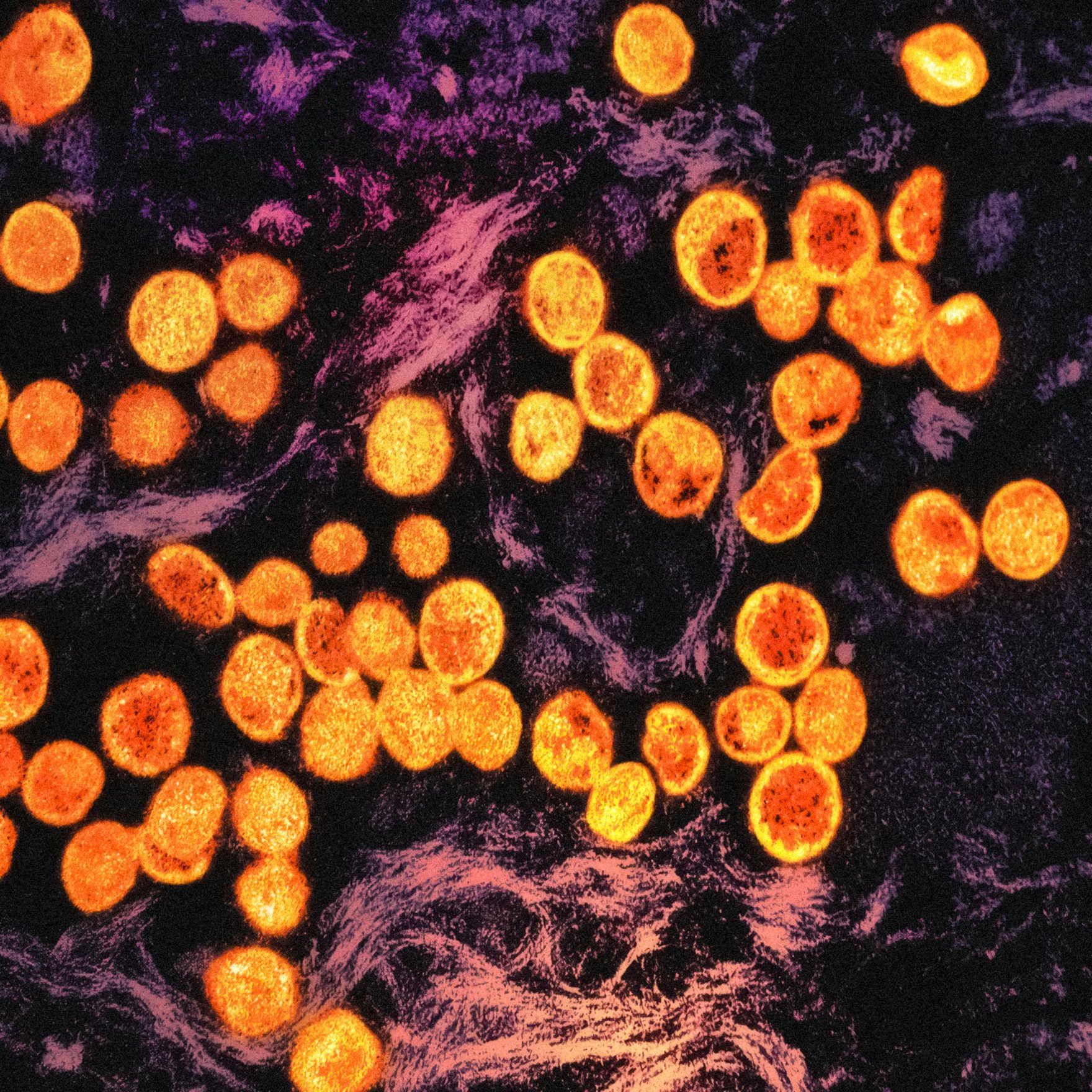
Produced by the National Institute of Allergy and Infectious Diseases (NIAID), this highly magnified, digitally colorized transmission electron microscopic (TEM) image highlights the particle envelope of a single, spherical shaped, Middle East respiratory syndrome coronavirus (mers-cov) virion, through the process of immunolabeling, the envelope proteins, using rabbit HCoV-EMC/2012 primary antibody, and goat anti-rabbit 10nm gold particles.
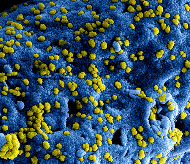
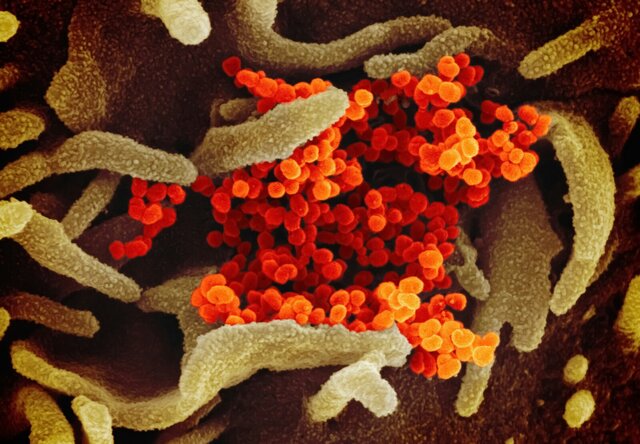
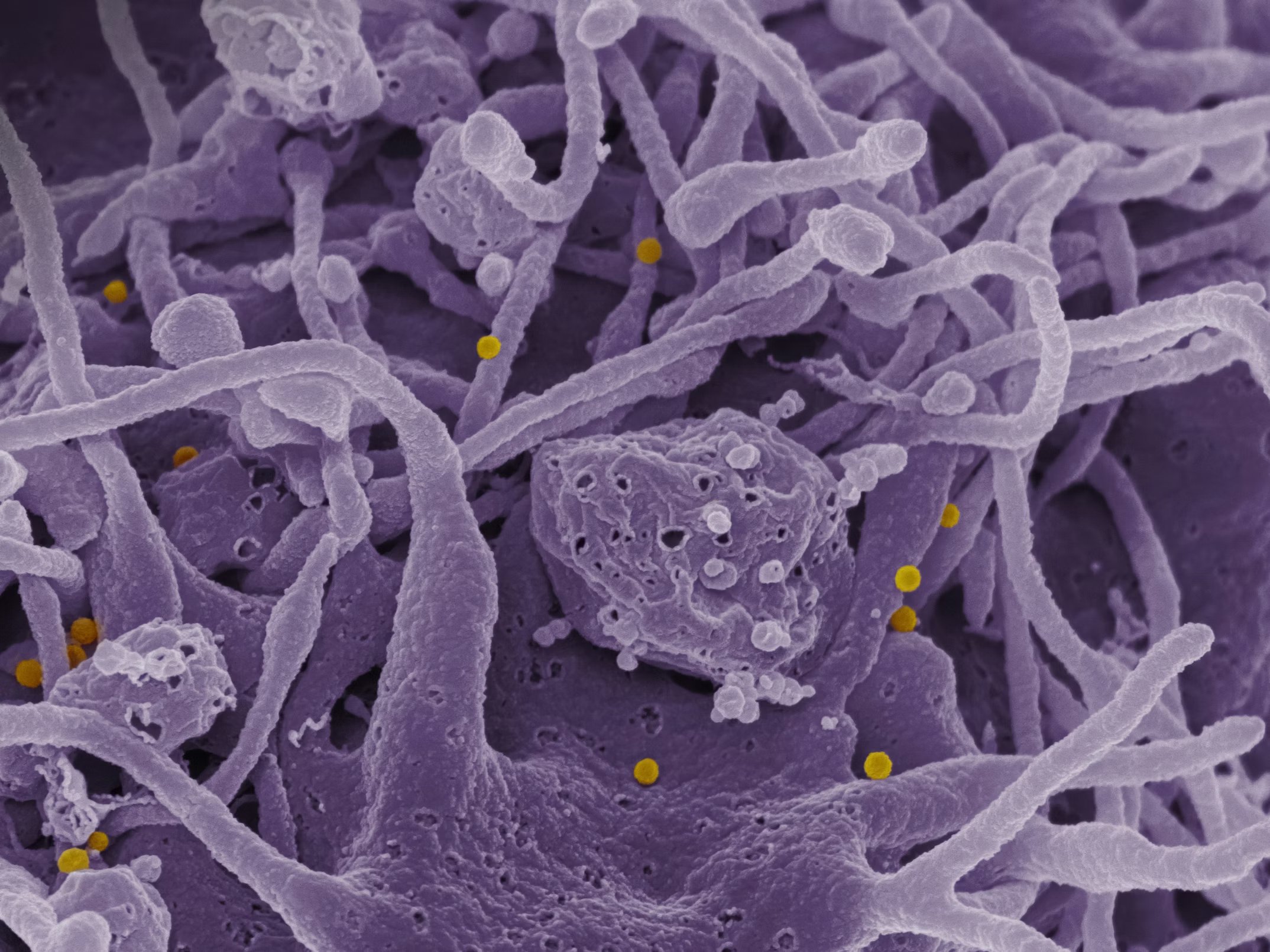
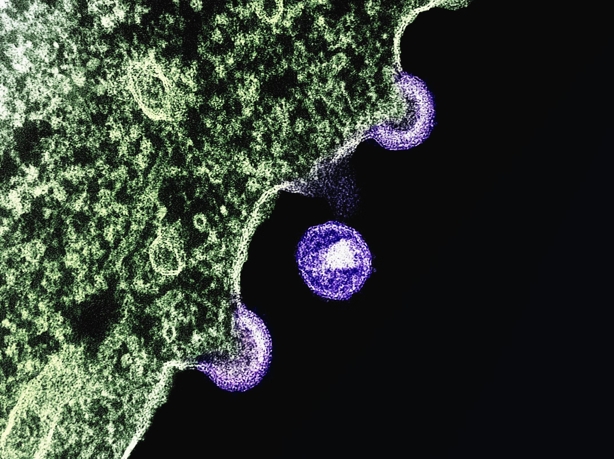
mers-cov
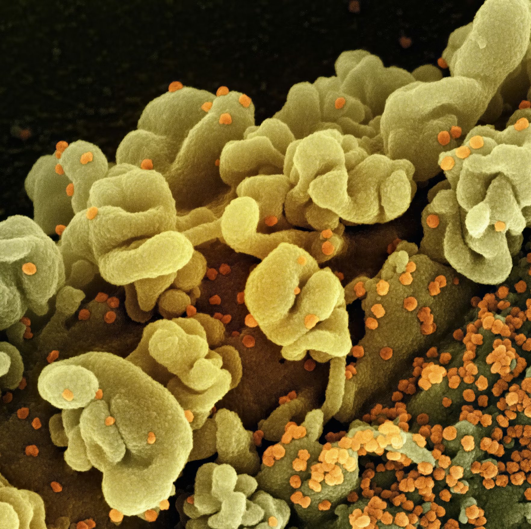
Produced by the National Institute of Allergy and Infectious Diseases (NIAID), this highly magnified, digitally colorized scanning electron microscopic (SEM) image, revealed ultrastructural details at the site of interaction of numerous yellow colored, Middle East respiratory syndrome coronavirus (mers-cov) viral particles, located on the surface of a Vero E6 cell, which had been colorized blue.


Crimean-Congo Hemorrhagic Fever (CCHF) Virus Scanning electron
Crimean-Congo Hemorrhagic Fever (CCHF) Virus Scanning electron micrograph of Crimean-Congo hemorrhagic fever (CCHF) viral particles (yellow) budding from the surface of cultured epithelial cells from a patient. Credit: NIAID https://www.flickr.com...
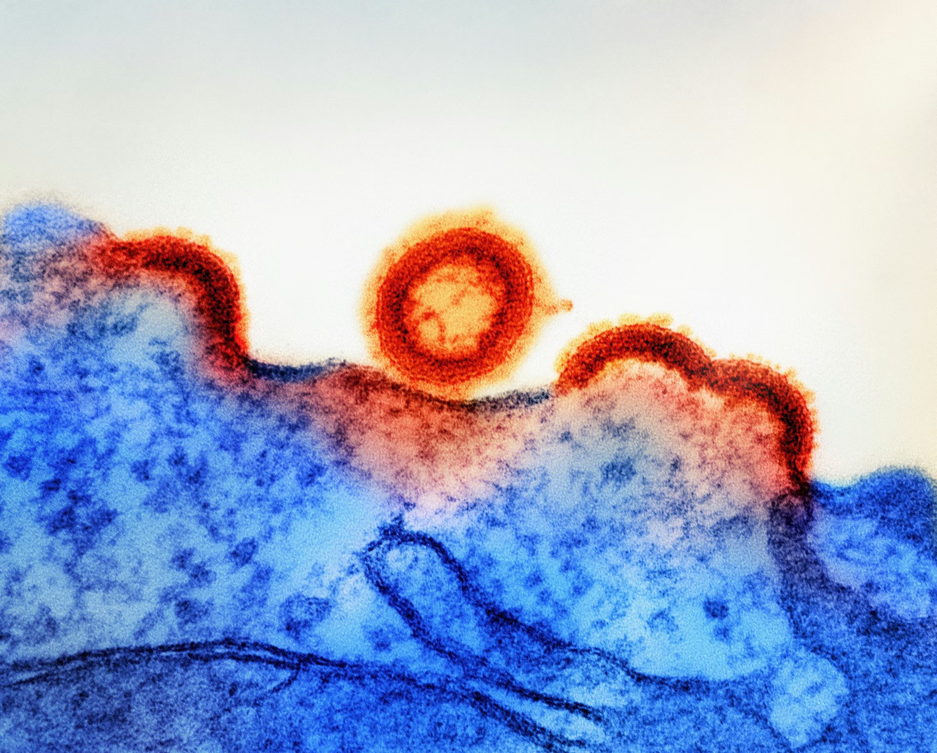
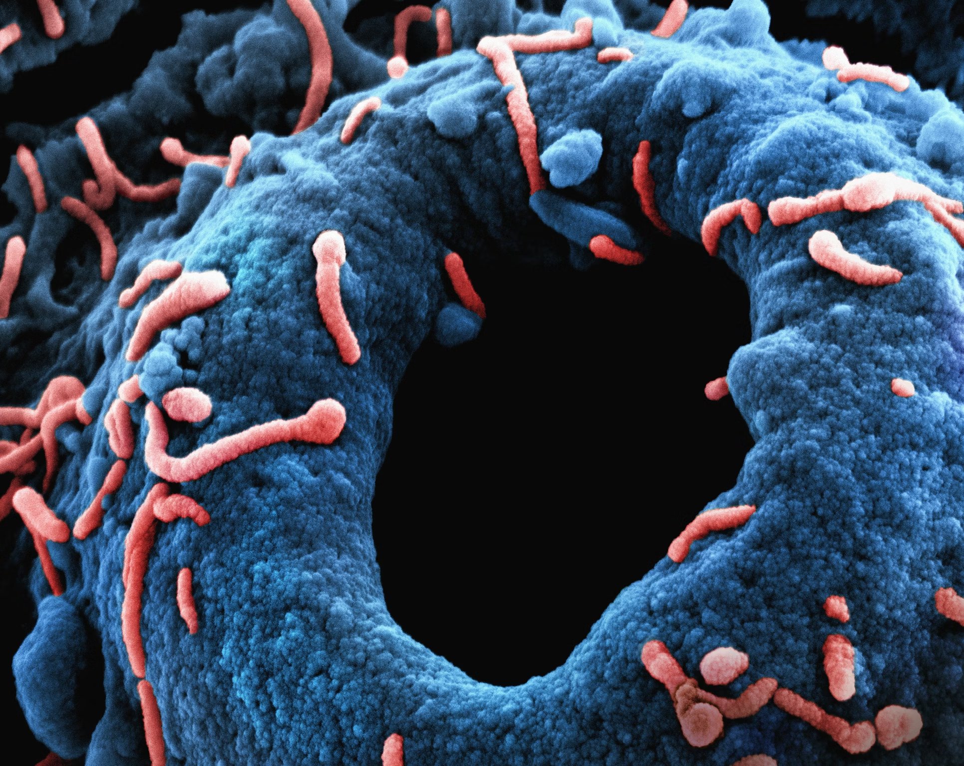
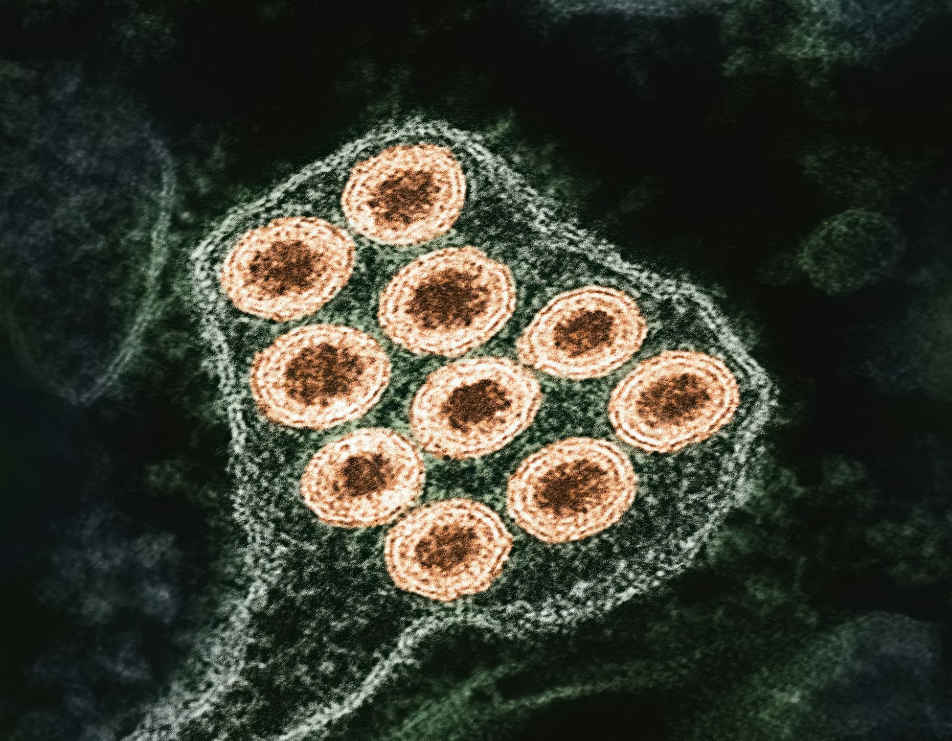
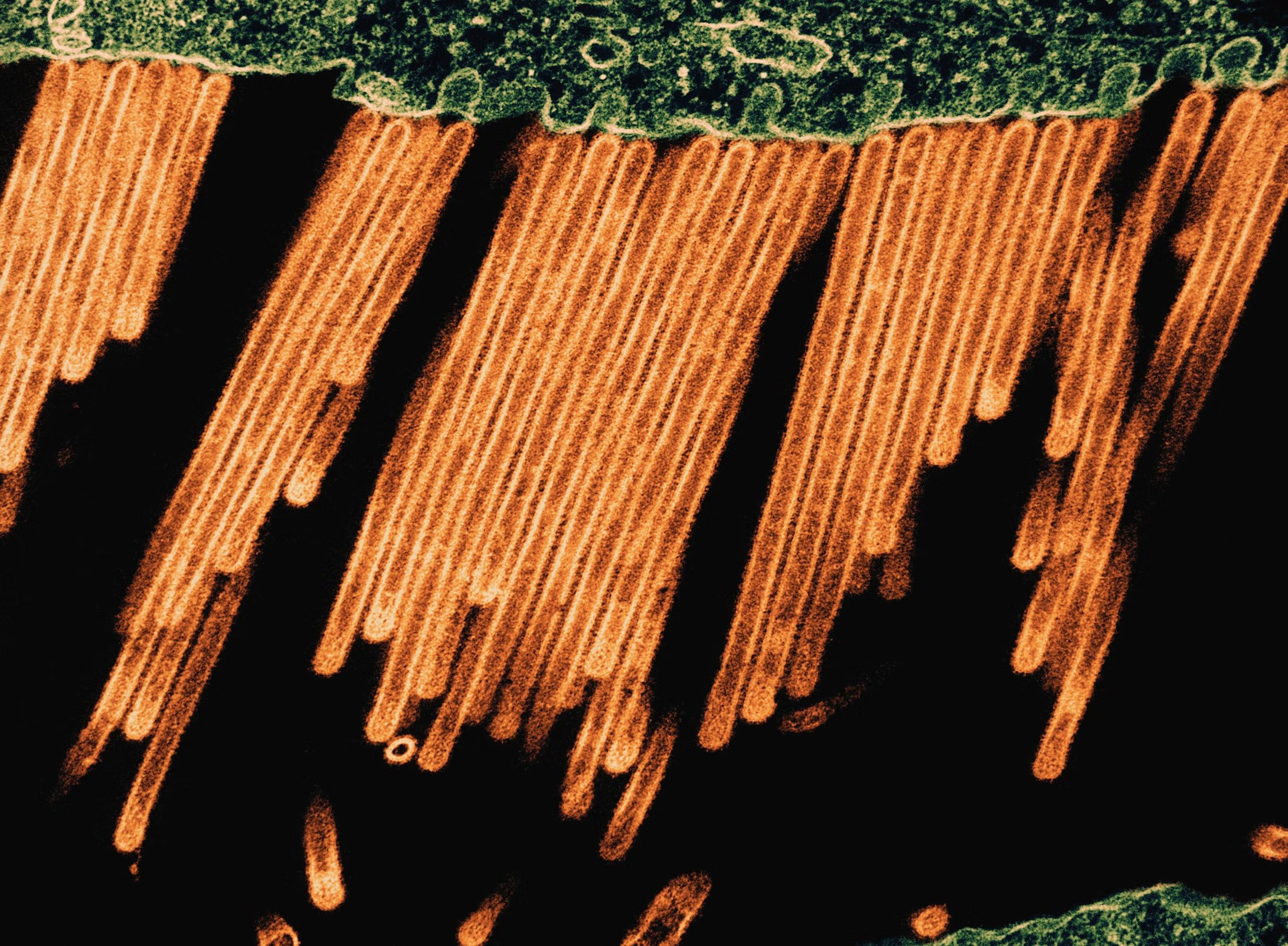
HIV-1 Virus Particles Transmission electron micrograph of HIV-1 virus particles
HIV-1 Virus Particles Transmission electron micrograph of HIV-1 virus particles (colorized red/yellow) replicating from an HIV-infected H9 T-cell (blue). Budding virus particles that have not yet separated from the cell appear as semi-circles. A s...

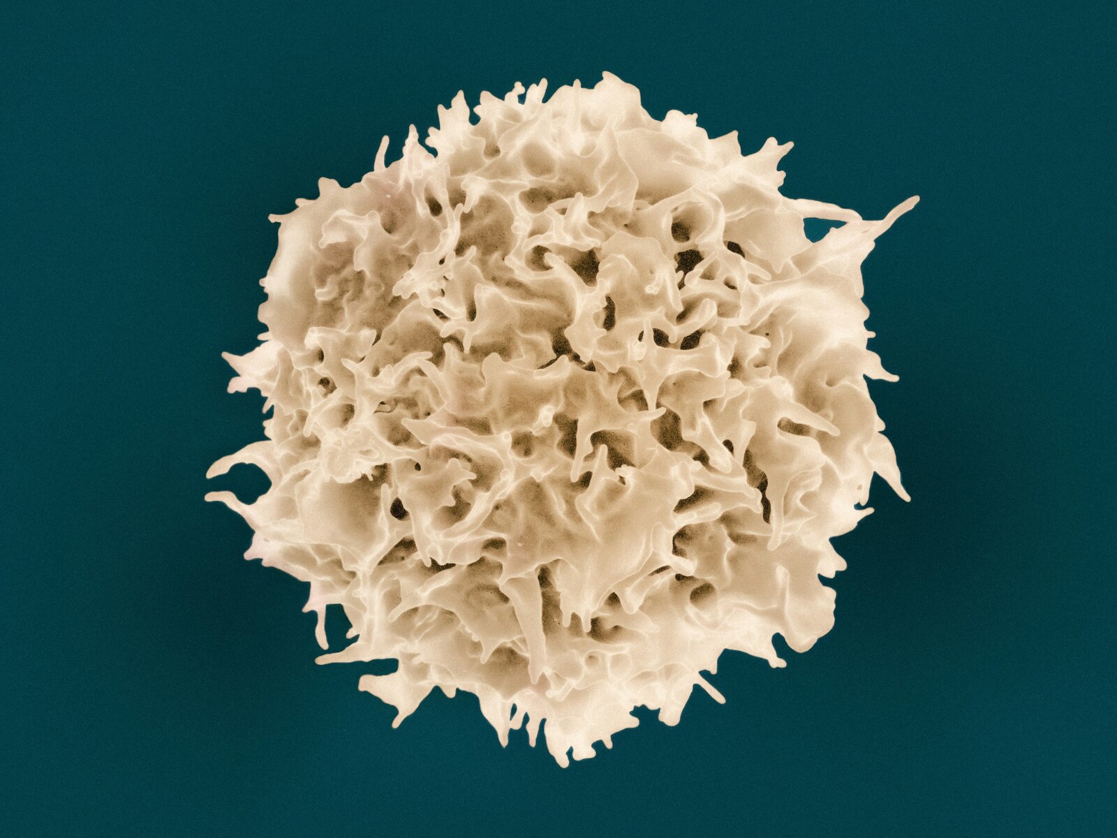
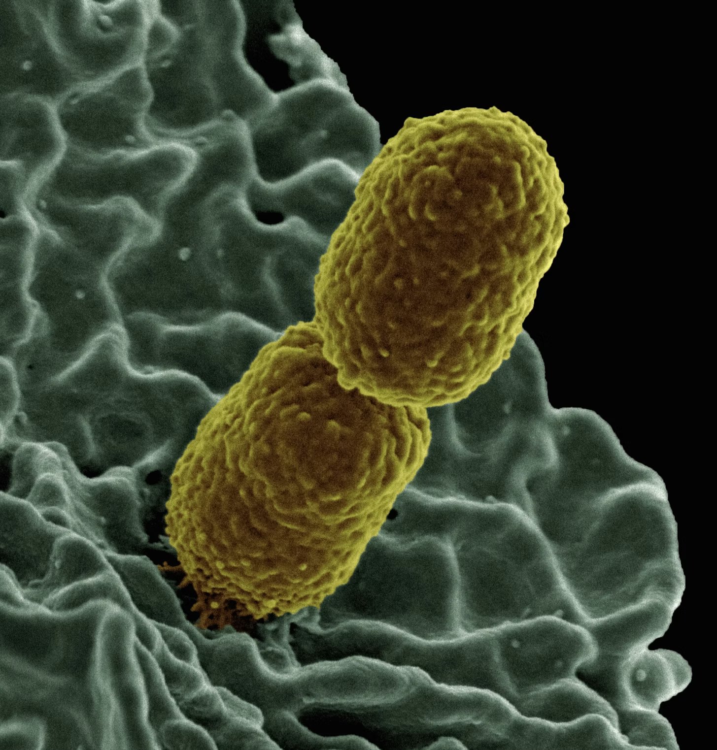
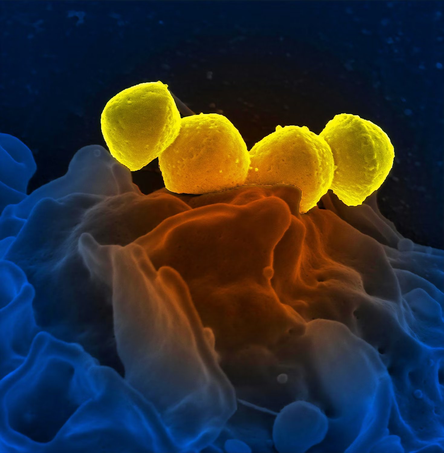
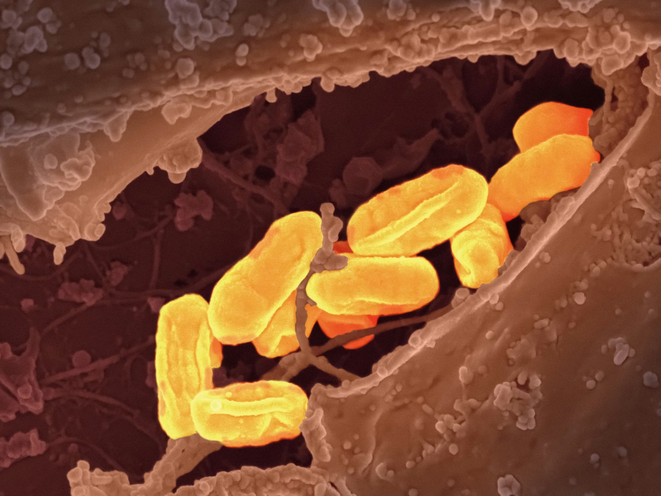
Methicillin-Resistant Staphylococcus aureus (mrsa) Bacteria
Methicillin-Resistant Staphylococcus aureus (mrsa) Bacteria Colorized scanning electron micrograph of methicillin-resistant Staphylococcus aureus (mrsa) bacteria purified from a cell culture laboratory sample. Image captured at the NIAID Integrate...


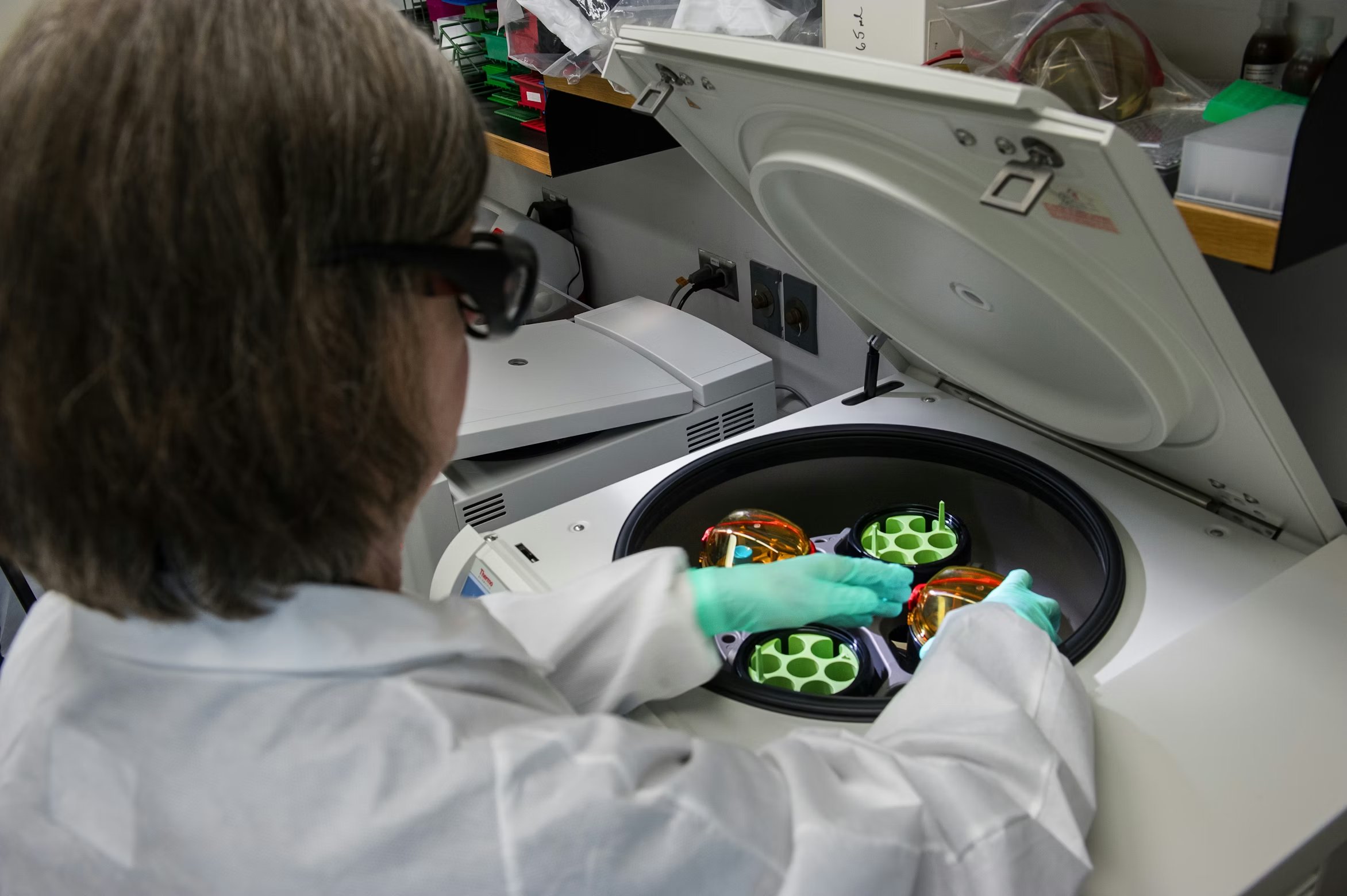
CDC scientist was shown working with stool samples
This Centers for Disease Control and Prevention (CDC) scientist was shown working with stool samples, which had been mixed with chemicals, and was placing the chemical mixtures into a centrifuge, which when spun up to a high rate of speed, would s...
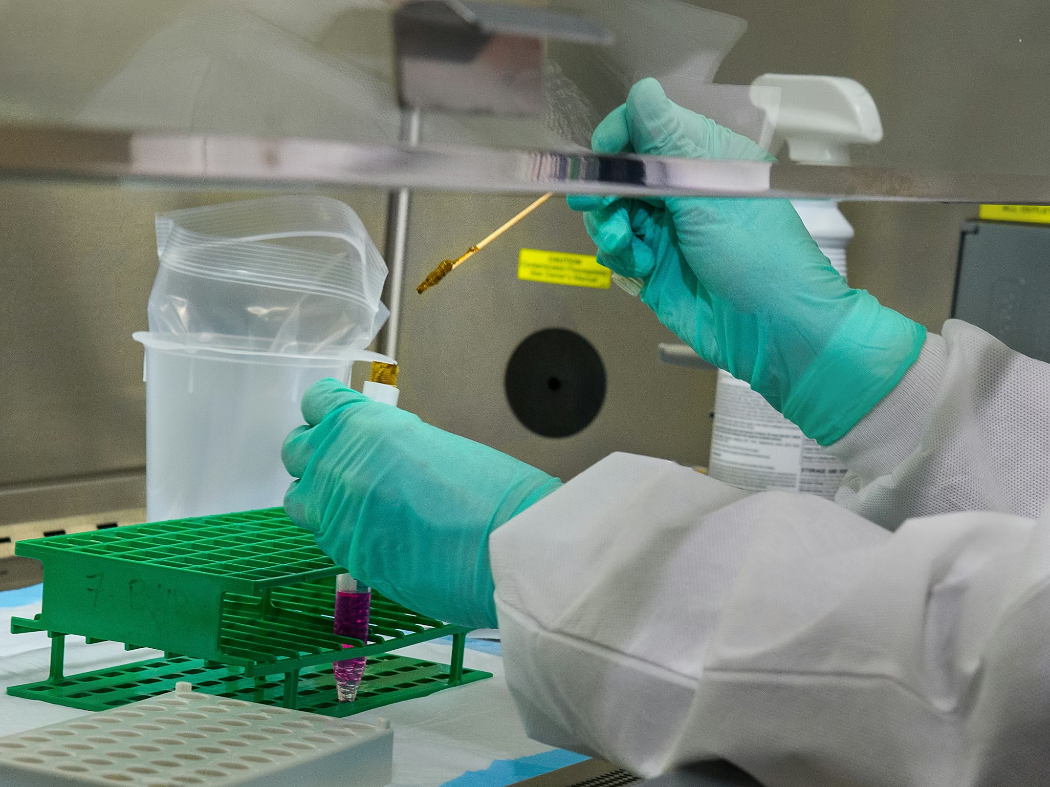
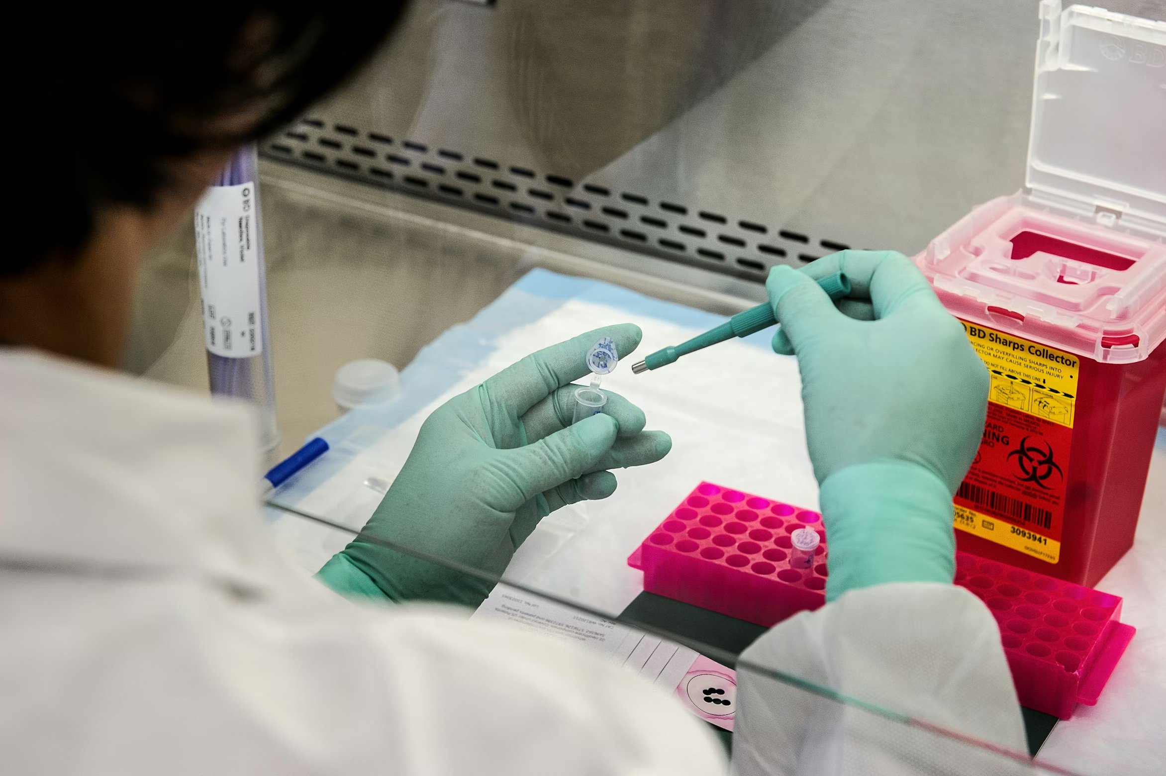
CDC scientist was shown adding a stool sample to the cell culture medium
To mix the stool and the chemicals together, this Centers for Disease Control and Prevention (CDC) scientist was shown adding a stool sample to the cell culture medium, along with glass beads, which will suspending the stool in the solution. Then ...
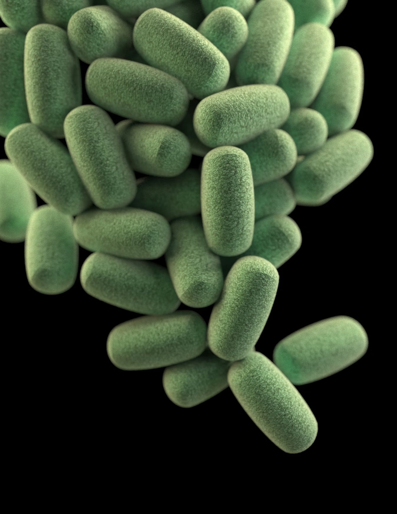
3D computer-generated image of a cluster of barrel-shaped, Clostridium perfringens bacteria
This illustration depicts a three-dimensional (3D), computer-generated image of a cluster of barrel-shaped, Clostridium perfringens bacteria. The artistic recreation was based upon scanning electron microscopic (SEM) imagery. See PHIL 21914, for a...
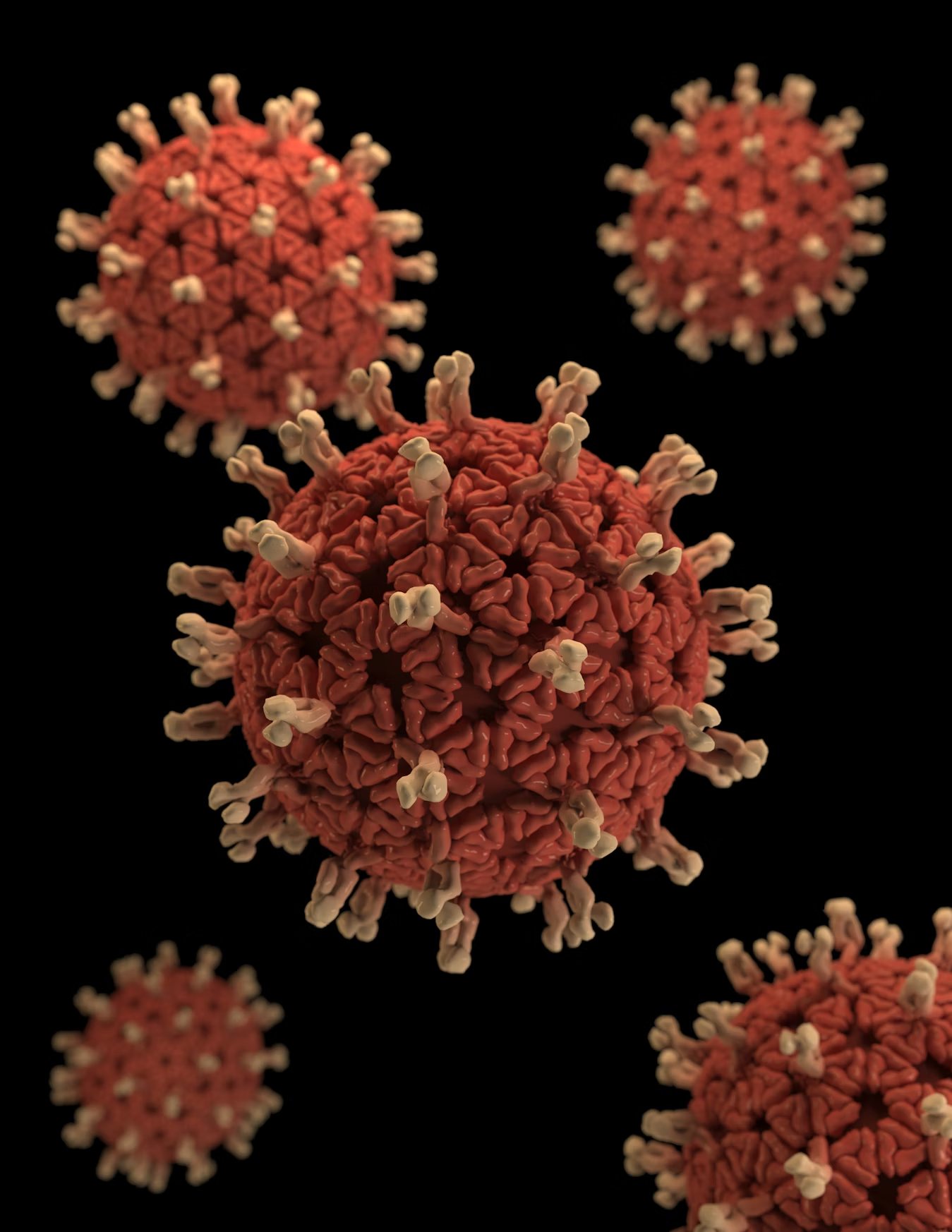
3D graphical representation of a number of Rotavirus virions
This illustration provided a 3D graphical representation of a number of Rotavirus virions, set against a black background. Note the organism's characteristic, wheel-like appearance, which was made visible when viewed under the electron microscope....
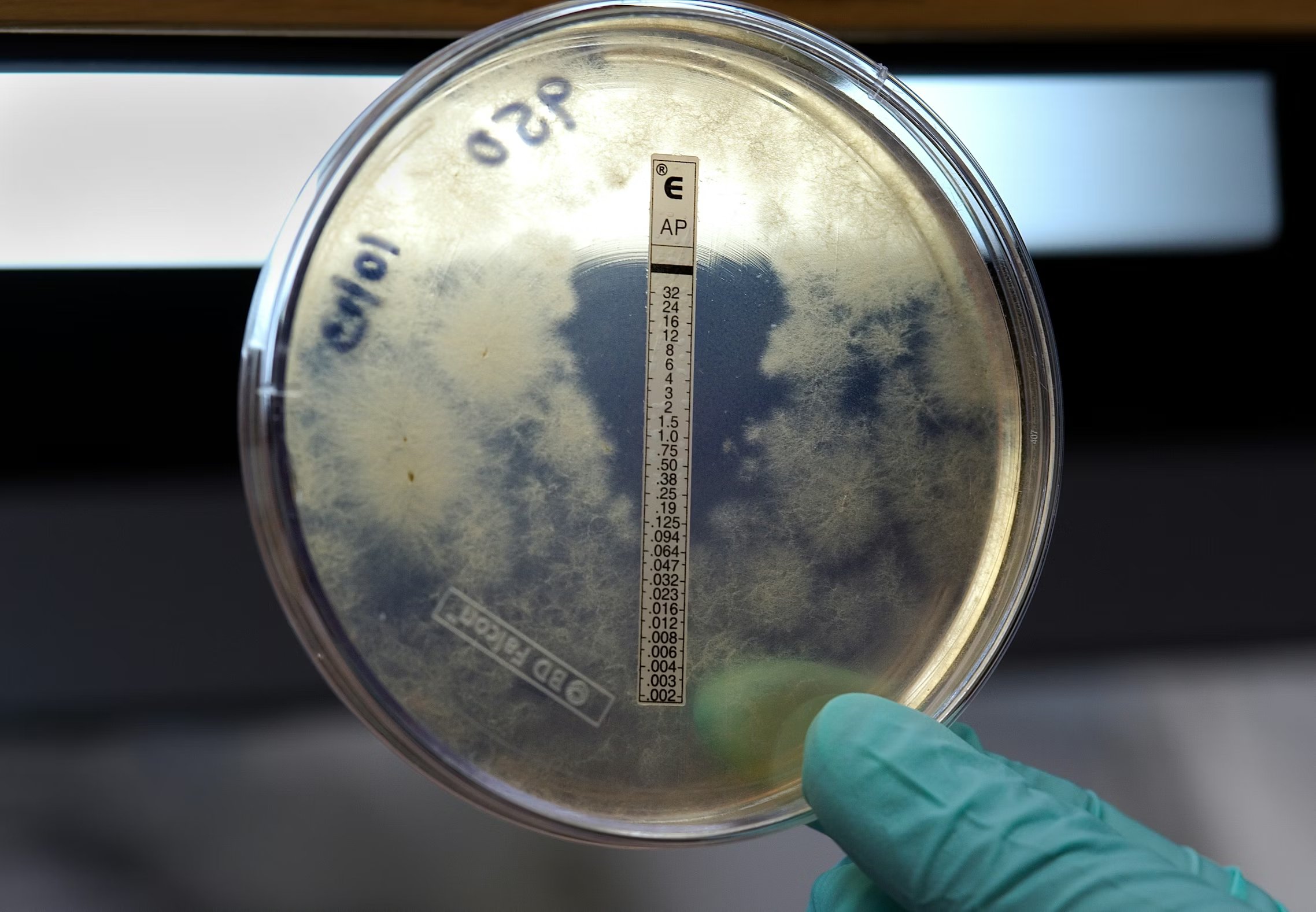
CDC susceptibility test to the antifungal drug, amphotericin B
Captured during the Centers for Disease Control and Prevention's (CDC) multistate meningitis outbreak investigation, this plate revealed the results of a susceptibility test to the antifungal drug, amphotericin B. The drug inhibited growth of the ...
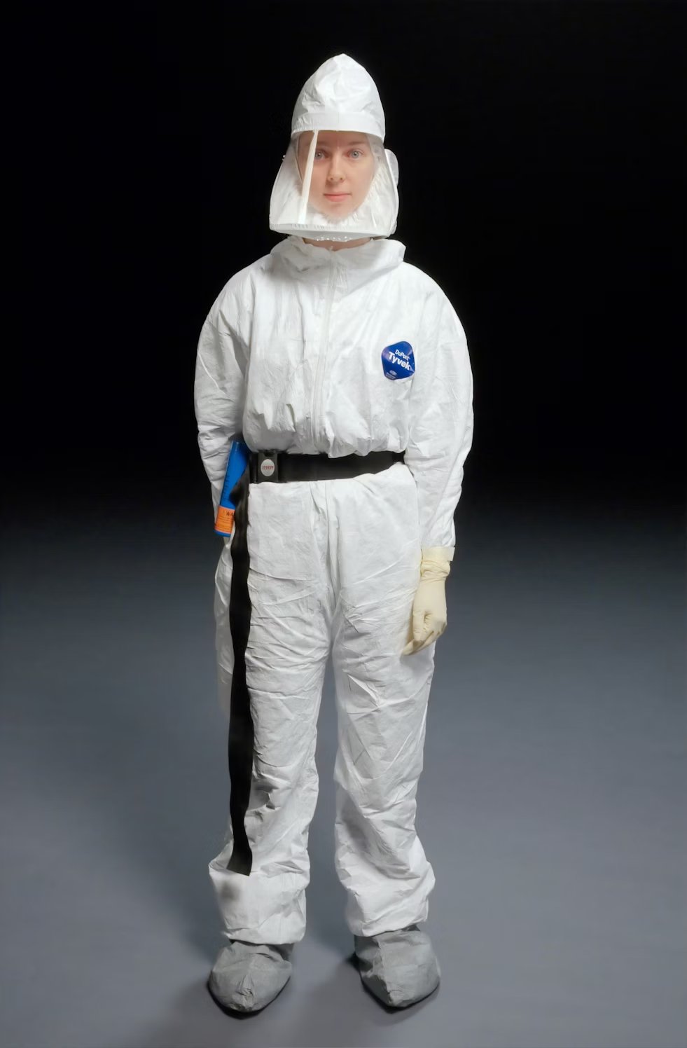
CDC scientist demonstrating how to wear personal protective equipment
This 2008 photograph depicted a Centers for Disease Control and Prevention (CDC) scientist demonstrating how one is to properly wear personal protective equipment (PPE) in appropriate laboratory settings. This PPE outfit included the Tyvek® suit, ...

Administering the H1N1 live attenuated intranasal vaccine (LAIV) to an Indian female recipient.
This 2009 image depicts a healthcare practitioner as he was administering the H1N1 live attenuated intranasal vaccine (LAIV) to an Indian female recipient. Using a small syringe, he was delivering the vaccine mist into the woman's right nostril. P...